NOT a cell with chromosomes pulling apart. Which image represents cytokinesis in a plant cell.
What Is The Difference Between Animal And Plant Cytokinesis Quora
- cytokinesis stock pictures.

. Cytokinesis in plant cells is formed through vesicles in the centre of a. 2 Show answers Another question on History. 3 MULTIPLE CHOICE OPTIONS.
Cytokinesis relies on a tight interplay between signaling and cellular mechanics and has attracted the attention of both biologists and physicists for more than a century. Spindle fibres microtubules and chromosomes are still visible. The 3rd image good luck Send.
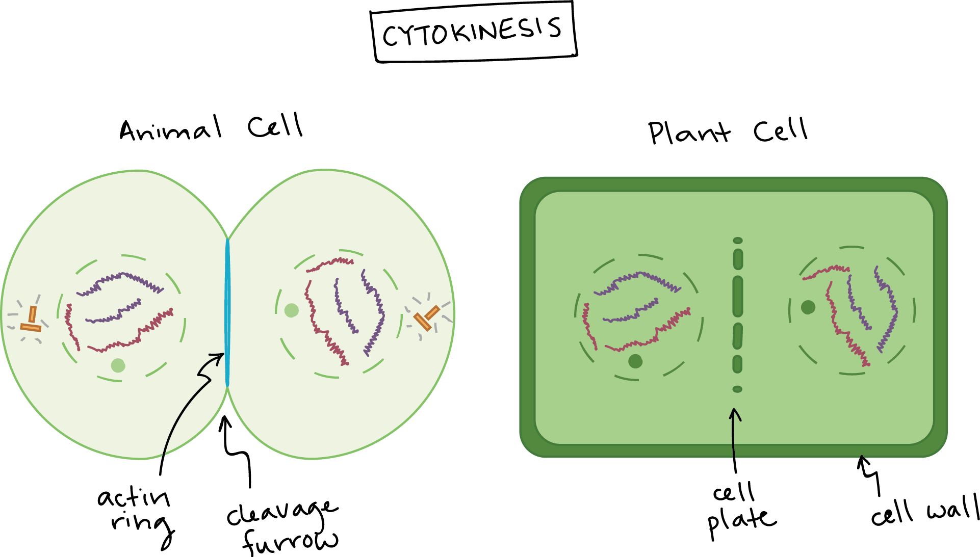
Which image represents cytokinesis in a plant cell. During cytokinesis the cytoplasm splits in two and the cell divides. Cytokinesis happens by division of the cytoplasm which occurs by the formation of cell plates in plants But when it comes to animal cells Cytokinesis occurs through cleavage.
Which part of the cell cycle does this image represents in an animal cell. Which image represents the step in mitosis when chromosomes condense and spindle fibers form. Which image represents cytokinesis in an animal cell.
Science - 4th. An animal cell left and a plant cell right are shown. Which describes what she should draw.
Report an issue. Which describes what she should draw. Which organelle labeled x in the diagram is found in both plant and animal cells.
Which image represents cytokinesis in a plant cell. Cytokinesis relies on a tight interplay between signaling and cellular mechanics and has attracted the attention of both biologists and physicists for more than a century. Now the main difference between cytokinesis in plant and animal cell is plant cells have a cell wall that needs to be split while animals do not have any cell wall.
Arthur made a mistake labeling the diagram of the three stages of the cell cycle. Plants and Animal Adaptations. Microtubules are depicted in blue the actomyosin contractile ring and the midbody ring in red and the endosomal sorting complex required for transport ESCRT-III spiral filaments in green.
In animal cells cytokinesis occurs through cortical remodeling orchestrated by the anaphase spindle. Cytokines is the third image. Jacqueline trying to draw an image of a cell in telophase.
Scanning electron micrograph of just-divided hela cells. The process of division of cytoplasm in the plant cell is called cytokinesis in plant cells whereas the process of division of cytoplasm in an animal cell is called cytokinesis in an animal cell. If a cell undergoes mitosis but does not complete cytokinesis it would become a cell with nuclei.
In plant cell cytokinesis cell plate formation takes place to divide cytoplasm into two daughter cells. You might be interested in. Prisoha 69 1 year ago.
The process is different in plant and animal cells as you can see in Figure PageIndex8. Biology 21062019 2000 ctyrector. Gospel of wealth ap us.
The cytokinesis process in the animal cell is attributed to the role of the contractile ring. A teacher makes a venn diagram. A contractile ring composed of actin filaments forms just inside the plasma membrane at the former metaphase plate.
In animal cells cytokinesis occurs through cortical remodeling orchestrated by the anaphase spindle. Label with the following terms. Cytokinesis the final step in cell division partitions the contents of a single cell into two.
The actin filaments pull the equator of the cell inward forming a. - In the animal cells the process initiated from anaphase and ends at telophase. Verdich 7 1 year ago.
Cytokinesis in plant cells forms cell walls whereas Cytokinesis in animal cells doesnt. Cleavage furrow cell plate Whitefish embryo Allium onion root tip 2. Observe a cell that represents cytokinesis in whitefish embryo and onion root tip to compare animal vs.
As animal cells that lack cell walls cytokinesis follows the onset of anaphase. Schematic diagram illustrating the different stages of cytokinesis in animal cells. - cytokinesis stock pictures royalty-free photos images.
Which are the main stages of the cell cycle. 1 MULTIPLE CHOICE OPTION. Which image represents cytokinesis in an animal cell.
Answerthe third image represents. Which step of mitosis involves the condesing of DNA into chromosomes. Which image represents cytokinesis in an animal cell.
Whos idea was it for businessmen to use their wealth for the greater good of society. Cytokinesis the final step in cell division partitions the contents of a single cell into two. Which image represents cytokinesis in an animal cell Other questions on the subject.
Which image represents cytokinesis in an animal cell. Whitefish mitosis whitefish embryo blastula telophase cytokinesis daughter cells magnification x250 cleavage furrow has constricted the cell into two daughter cells. A schematic representation showing the reorganization of an animal cell progressing through the different stages of cytokinesis.
In animal cell a cleavage is formed first of all in the middle of the cell that has to be divided the cleavage deepens until it meets the membrane and then eventually cell. How has urban renewal affected poorer. Examine the images of a plant cell in the different stages of mitosis.
In animal cells the plasma membrane of the parent cell pinches inward. Jacqueline is trying to draw an image of a cell in telophase.

1 Which Letter Represents Mitosis And Cytokinesis Ppt Download

File Plant And Animal Cell Cytokinesis Svg Wikipedia
Phases Of Mitosis Mitosis Biology Article Khan Academy

Mitosis Meiosis Hw Flashcards Quizlet

Which Image Represents Cytokinesis In An Animal Cell Brainly In
Which Image Represents Cytokinesis In An Animal Cell Brainly In

Which Image Represents Cytokinesis In An Animal Cell Brainly In

Which Image Represents Cytokinesis In An Animal Cell Brainly In
0 comments
Post a Comment